
“Even after the light fades, I still see it... and the words—I hear them again.”
Introduction
A 19-year-old girl walked into our Neurology OPD with complaints of recurrent throbbing headaches, predominantly frontal and right-sided, often accompanied by nausea and vomiting.
No trauma. No medical history.
But the headache was just the beginning.
Upon taking the history further, she described something that made both me and the senior consultant pause :
“I see images even after they’re gone. And I hear the same sound twice or more in the opposite ear”
Palinopsia: When Sight Lingers
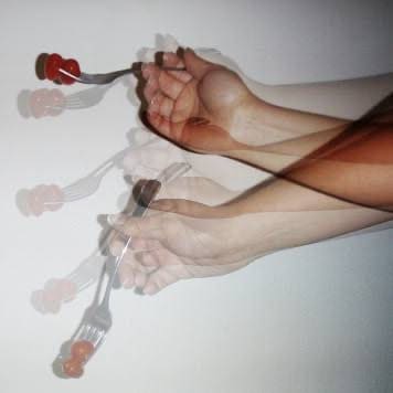
Palinopsia is a rare and often misunderstood visual disturbance in which images persist or reappear even after the original object is no longer in view.
It is broadly classified into two subtypes:
- Illusory Palinopsia: A visual persistence or afterimage, often called a fading trail or motion smear, is something you can see when you look at bright things or in low light.
- Hallucinatory Palinopsia: More vivid and detailed visual replays—seeing fully formed images that were previously present but are no longer in view, almost like a mental “photograph” projected back into sight.
The underlying causes are varied and span several domains:
- Neurological: Occipital or parietal lobe lesions, seizures, migraines
- Psychiatric: Visual hallucinations in mood or psychotic disorders
- Pharmacological: Side effects of medications such as topiramate, trazodone, or hallucinogens
In her case, she vividly described that any object she focused on would leave behind a ghost-like trail, lingering for several seconds before fading, whether it was a person walking by, a bright light, or even her hand.
It was as if her eyes had become a projector, replaying scenes that had already passed.
This illusion of time-lagged vision pointed toward a dysfunction in her visual processing pathway, later localised to the right temporo-parietal region—a critical hub for integrating spatial and sensory information.
Palinopsia is more than just an afterimage—it’s the brain’s way of clinging to visual inputs, sometimes as a warning that something deeper is wrong.
Palinacousis: When Sound Echoes Again
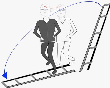
Even more peculiar than her visual symptoms was her description of an auditory echo—not just a reverberation, but a distinct repetition of sound in the opposite ear, several seconds after the original.
“If someone spoke into my right ear, I’d hear the exact same thing echo from the left, 3–5 seconds later.”
This phenomenon is known as palinacousis—a rare auditory disturbance where sounds are perceived again after the original stimulus has ended, typically in the absence of any actual external repetition.
In contrast to tinnitus or hallucinations, palinacousis entails the precise reproduction of actual environmental sounds; patients frequently refer to these as delayed echoes or auditory “afterimages.”
It is most frequently linked to dysfunction of the temporal lobes, particularly the auditory cortex, and can be brought on by:
- Structural brain lesions
- Seizure activity
- Tumours
- Stroke
- Rarely, post-infectious or inflammatory conditions
In her case, this symptom helped localise the pathology to the right temporo-parietal region, later confirmed on imaging.
Palinacousis may seem like a benign quirk—but as this case shows, it can be a subtle clue to more profound neurological disruption.
Investigations and Diagnosis
Despite these strange symptoms, her vitals were stable, and there were no neurological deficits on examination. She had no seizure, no motor or sensory complaints.
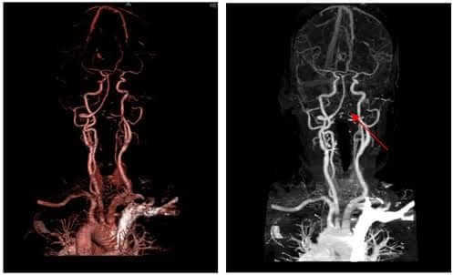
A detailed clinical history is essential in diagnosing Palinopsia, along with a neurologic examination. Diagnostic imaging (MRI or CT) may be required to identify structural brain abnormalities.
Electroencephalography (EEG) might be used if seizures are suspected. Additionally, a thorough review of any medications or substance use is crucial.
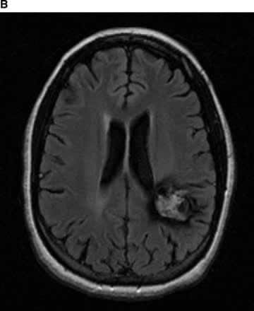
After taking the history and ruling out various causes of the symptoms, our senior consultant advised an MRI Brain, which revealed a small Astrocytoma in the right temporo-parietal region, an area critical for both visual and auditory processing.
Management
Given the tumour’s small size and stable symptoms:
- Medical management was initiated
- Regular follow-ups and neuroimaging scheduled
- Surgical resection was to be considered if symptoms progressed
Symptoms determine the type of treatment:
- CBT (cognitive behavioural therapy) for psychological coping
- Anticonvulsants for seizures
- Management of migraines
Lessons Learned
Although they may seem unreal, Palinopsia and palinacousis are real and upsetting. They may indicate actual neurological pathology, but are frequently written off as anxiety or “all in the head.”
A precise diagnosis necessitates:
- Neuroimaging
- Careful history
- High index of suspicion
An Expression of Gratitude
When I was given this case, I was a neurology medical officer. I am very grateful to my consultant for his patience and wisdom. His explanation showed me how subtle sensory symptoms can reveal profound neurological truths.
Concluding Thoughts
When the brain sends after images or echoes, it might be trying to communicate a deeper meaning.
We need to pay attention to our patients’ stories as well as their symptoms.
Because sometimes a patient’s story can provide more diagnostic information than a scan.

